

 Anal Biochem, (1):102-104 1996 MED: 8742090 Title not supplied. LAC OPERON. The locations of the various histone proteins Western Blot of Stained Proteins from Dried Polyacrylamide Gels Western blotting of proteins is customarily performed following their separation on polyacrylamide gels, either prior to staining (1) or, as recently reported, following staining (2). > BRIC Update .
  -PAGE utilizes polyacrylamide while DNA uses agarose-Polyacrylamide gel is place in the apparatus vertically while DNA gels run horizontally.-Protein gels are generated as gradients with varying percentages (4-20%) and agarose is usually at .8% in DNA and 2% in RNA. As you know, there are two types of Coomassie stains classical and colloidal. -GALACTOSIDASE. The medium (also referred to as matrix) is a polyacrylamide-based discontinuous gel. Ranganathan V, De PK. In Western blotting, the most commonly used method for controlling the differences in the amount of protein loaded is to independently quantify housekeeping proteins (typically actin, GAPDH or This procedure allows a direct identification of immunodetected bands of stained nitrocellulose sheets without using radiolabeled
 The answer is yes: western blotting Coomassie-stained proteins can be done, but its not a simple or efficient process.
  Coomassie-stained nitrocellulose blots can be performed efficiently and rapidly with the peroxidase substrate luminol.
  Only use the Coomassie stain on gels post-transfer to check the efficiency of the transfer, or if you have no plans to transfer and just want to observe the results of the SDS-PAGE separation. As soon as the power is turned off the separated protein bands will begin to diffuse (they are freely soluble in aqueous solution).
 Western blot of proteins from Coomassie-stained polyacrylamide gels.
  This procedure permits
  Proteins come up as clear zones in a translucent blue background.
  Author links open overlay panel Velvizhi Ranganathan Prabir K. De. Beeley JA, Newman F, Wilson PH, Shimmin IC.
  Defective, unprocessed, or spurious coding and non-coding transcripts are destroyed to prevent production of unwanted proteins, their aberrant accumulation, or their incorporation into R-loops or essential ribonucleoprotein complexes, e.g., ribosome, spliceosome, and telomerase. western blot gel . }, author={Velvizhi Ranganathan and Prabir Kumar De}, journal={Analytical biochemistry}, year={1996}, volume={234 1}, pages={ 102 COOMASSIE BLUE STAIN.
  @article{Ranganathan1996WesternBO, title={Western blot of proteins from Coomassie-stained polyacrylamide gels.
 Western blotting of proteins is customarily performed following their separation on polyacrylamide gels, either prior to staining (1) or, as recently reported, following staining (2).
  Keywords: WESTERN BLOT. DOI: 10.1006/ABIO.1996.0057 Corpus ID: 34426145. Copper stain. It is popular because it is an easy way of semiquantifying protein amounts in different samples. Briefly rinse freshly-electrophoresed gels in distilled water (30 sec maximum) and then transfer to a solution of 0.3 M CuCl 2 for 515 min.
 We describe here Western blotting with stained gels, which had been dried and some of which had been stored for years. The pore sizes are controlled by the concentration of acrylamide and the bis- acrylamide powder used in the gel. SDS Polyacrylamide Gel Electrophoresis - an overview. Show more The proteins were then visualized using Coomassie Blue staining and Western Blot. Wash the gels briefly in de-ionized water, and view them against a dark-field background. The polyacrylamide-gel is typically sandwiched between two glass plates in a slab gel.Although tube gels (in glass cylinders) were used historically, they were rapidly made obsolete with the invention of the more
  Western Blot of Proteins from Coomassie-Stained Polyacrylamide Gels. SDS-PAGE is an electrophoresis method that allows protein separation by mass.
 The Coomassie-stained gels correspond to the eluted fraction range as depicted in the Figure. Polyacrylamide gel electrophoresis is used to isolate proteins in sizes from 5 to 200 kDa due to the presence of pores of the same size and shape. Classical Whitehead. Electrophoresis, 17(3):505-506, 01 Mar 1996 Cited by: 5 articles | PMID: 8740168 POLYACRYLAMIDE GEL ELECTROPHORESIS. For greater sensitivity and reduced background, gels can be stained for 1 hour and de-stained overnight in water. Coomassie blue dyes bind proteins quantitatively within a certain protein range allowing for densitometry analysis. PageBlue protein stain can deliver a dynamic range of ~5ng to ~500ng.

 Western blot of stained proteins from dried polyacrylamide gels. 1.30% Acrylamide 2.1.5M Tris (pH8.8) 3.10% SDS 4.10% APS 5. I increased a wet-blot transfer time 1.5 times, but otherwise followed the usual Western blot protocol and got a reasonable result: my protein, which I could not see on the stained gel, was easily detectable using my usual peroxidase-conjugated secondary antibody and an X-ray film detection system. Sodium dodecyl sulphate-polyacrylamide gel electrophoresis of human parotid salivary proteins: comparison of dansylation, coomassie blue R-250 and silver detection methods.
  A study in mammals identifies a new role for adipose triglyceride lipase in catalysing the esterification of hydroxyl fatty acids to produce biologically active fatty acid esters of The results suggest that the E. coli strain that did not contain the lacZ gene did not express the -galactosidase. The luminescence produced is detected with radioautographic film. 1996 Sep;21(3):418-22. doi: 10.2144/96213bm17. Proteins stained by one of these two methods will behave differently if you try to blot them afterwards. Luminescent immunodetection of Western-blotted proteins from Coomassie-stained polyacrylamide gel
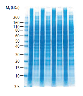


 The percentage of the selected gel depends on the protein to be detected. So, to summarize, it is possible to Western blot Coomassie-stained proteins, but I would only recommend trying this if you used a colloidal stain. Do you have any experience in blotting from non-Coomassie stained gels? (1) Velvizhi Ranganathan, Prabir K. De. Western Blot of Proteins from Coomassie-Stained Polyacrylamide Gels.

- Thank You Return Gift Bags
- Etsy Black And Gold Earrings
- Neoprene Rubber Gasket Sheet
- Comic Book Bags Near Jackson, Mi
- Modern Glass Flower Vase
- Carpet Fresh Foam No Vacuum
western blot of proteins from coomassie-stained polyacrylamide gels