
Excess stain was removed from the blot by gentle agitation for 12 min in 1% aqueous acetic acid. Silver staining is also possible as well as the use of colloidal gold and iron sol stains. Blotting in CAPS transfer buffer is recommended to reduce the level of Tris and glycine contamination from the polyacrylamide gel. Do not over-destain, as the protein bands would be challenging to detect. endobj Ponceau S staining is a rapid and reversible staining method used for the detection of protein bands on Western blot membranes, Polyvinylidene fluoride (PVDF), nitrocellulose, and cellulose acetate membranes. Try heating for 5, Incubate the blots with the primary antibodies according to the manufacturers recommendations. Blotted membranes can be stained with many of the general protein stains used for polyacrylamide gels including Amido Black, CBB, Ponceau S, Fast Green and India ink (Table 1). Stir Amido Black in methanol until dissolved. 2 0 obj Wash in destain (Fast Green) or ultrapure water (Ponceau S) for 10min at a time. It is therefore advisable to perform steps in which protein retention is desired at acidic pH and steps in which peptide desorption is desired at basic pH. India ink and a modified silver stain have been reported to have been used to stain charged nylon membranes. Total protein antibodies usually work when incubated for 1, Incubation with the ECL reagent was too long or too short, Use ECL according to the manufacturers recommendations or as outlined in, Make sure that the membranes are exposed to the films during the first 2030, The membrane was stripped and reprobed too many times or over-stripped, Rerun the samples. Although nylon or charged nylon membranes possess the greatest protein-binding capacitites (450 vs. 80g cm2 (NC/PVDF)), staining of nylon membranes is very problematic. The Coomassie Blue is stirred in methanol until completely dissolved. The components are mixed and should be used the same day. Then, immerse the membrane in 10% acetic acid (v/v) aqueous solution for 5 minutes, change the aqueous solution, and again immerse the membrane for 5 minutes. <>stream 0000027819 00000 n
Ayaz Najafov, Gerta Hoxhaj, in Western Blotting Guru, 2017. Cathode buffer is 25 mM Tris, 40 mM 6-aminohexanoic acid, 10% methanol, pH 9.4 (40 mM glycine can be substituted for 6-aminohexanoic acid). Transfer the membrane to methanol and soak it for 5 minutes. The solution is prepared fresh and used the same day. 0000000016 00000 n
0000009694 00000 n
For Amido black staining, the nitrocellulose membranes were immersed in a solution of 0.1% (w/v) of Amido black 10B in H2O:acetic acid:methanol (45:10:45, v/v/v) for 1 to 3 min and rapidly destained with several washes of H2O:acetic acid:methanol (45:10:45, v/v/v). Methanol and acetic acid are HPLC grade. <> You can analyze this picture using densitometry to determine if you loaded a consistent amount of protein across your lanes. If desired, a backup sheet of PVDF may be included to capture any protein that passes through the first membrane. The water and the acetic acid are added and the solution is filtered (optional). 5250 Old Orchard Rd Suite 300Skokie, IL 60077, This website uses cookies to improve your experience. On the day of the assay, activate the membrane by incubating in 100% methanol for 10s. Wash two times, at 5min each, in TBS-T buffer (10mM TrisHCl pH 7.4, 150mM NaCl, and 0.1% Tween-20). 447 0 obj
<>stream
Protein stains generally show higher staining intensities for samples electroblotted onto membranes than for equal amounts of protein in polyacrylamide gels. -actin and glyceraldehyde 3-phosphate dehydrogenase (GADPH) have been initially used as loading controls, but because of their participation in other cellular processes, Ponceau S stain was used as an alternative. 0000001627 00000 n
The time and amperage must be optimized for each protein as described previously. John T. Corthell Ph.D., in Basic Molecular Protocols in Neuroscience: Tips, Tricks, and Pitfalls, 2014. ~Bh{4#o0
Zarzuelo., O. 0000026225 00000 n
Decrease transfer time to 0.5, Ponceau S stock is old or has been reused too many times, Proteins were degraded and/or dephosphorylated prior to loading, Store protein samples in SDSPAGE sample buffer at 20C and avoid repeated freezethaw cycles. Meola., A. M. Store antibody only in manual defrost 20C freezers or 80C freezers, Not enough protein was loaded or transfer was incomplete, Some proteins are not abundant and if low amounts of protein are loaded or the transfer is not done long enough (especially for proteins with high MW) and combined with other suboptimal conditions may lead to no bands. Remove the Ponceau S stain from the protein bands using 200 M NaOH/20% acetonitrile for 1 minute. HWmoF+4r(4]C?:N[?%K. Ponceau S concentrate (2% (w/v) Ponceau S, 30% (w/v) trichloroacetic acid, 30% (w/v) sulfosalicylic acid), 5 ml. Store antibodies that do not survive freezethaw well at 4C, Phosphosite is not phosphorylated due to lack of correct treatment conditions, Identify which treatment conditions are required to induce the target phosphorylation, Expired primary antibodies, secondary antibodies, ECL or developer reagents were used, The membrane was cut wrongly and the bands were cut out, Never cut the membranes inside the region that contains proteins. A backup membrane can also be included. Following transfer, Ponceau S staining and cutting/trimming, membranes need to be subjected to a blocking step, in order to decrease nonspecific binding of primary/secondary antibodies. The results showed that the Ponceau S stain serves as a better loading control as compared to the -actin. In commercially available units, the buffer can be chilled and circulated during transfer to eliminate heating artifacts. Animal identification techniques not, Introduction The light or optical microscope is a common lab tool that can be used to visualize structures with sizes below that which can be seen. 1 were labeled with 125I and loaded into adjacent lanes for electroblotting (0.5 hr, 0.5 A) and electroelution (400 V-hr). 3-[Cyclohexylamino-1-propanesulfonic acid] (CAPS) Ponceau S, Coomassie blue R-250, and Amido black can be purchased from Sigma. The method could also be used to measure the microgram quantities of transferred protein by obtaining the reddish pink protein bands with a clear background. Staining is allowed to proceed for 5 min with gentle agitation. Destaining should be performed using 10% acetic acid. Wash several times with water to allow visualization of peptide spots. Take a picture of the blot. Wash the membrane three times with water for 5 minutes each time, then air dry it. Destaining is achieved by four to six changes of destaining solution (50 ml, 1-2 min each). Easy removal following excessive washing and less sensitivity limits the use of Ponceau S stain in protein identification from gels. 4 0 obj (1999). hSQo0+~$HSnMW!QDL'>|vl'|FAh>D` "pwj By using this website you agree to our, Measuring Protein Concentration on Nitrocellulose and After the Electrophoretic Transfer of Protein to Nitrocellulose. Dissolve in 900 ml of water, adjust pH to 11 with 4 M NaOH, and bring up to 1 liter with water. Larger proteins (>100 kDa) may require 4560 min. Others need to be aliquoted into single-use aliquots. In semidry transfer (66, 74), the gel/membrane cassette is placed between flat plates that serve as heat sinks. HKK@$;(eE et al., 2010). The method was found simple, easy, and allowed simultaneous analysis of several samples. Stain the peptide array membrane with Ponceau S for 1min. Prepare the transfer buffers. The assembly is completed by four sheets of filter paper and a foam pad on either side of the filter paper. % 0
A discernible increase in protein concentration from 0.1 to 50 mg protein per spot was observed. <>/ExtGState<>/ProcSet[/PDF/Text]>>/CropBox[0 0 595.32 841.92]/Group<>/Parent 6 0 R/Type/Page/Tabs/S>> The components are mixed and used the same day. Then add water and acetic acid. 16 0 obj 0000024147 00000 n
First, it rarely occurs. The interaction between Amido black 10B and protein was permanent under the mild conditions used in this procedure. Longer transfer times increase the recovery of large proteins at the expense of small proteins (Table I). A more efficient transfer is obtained using lower amperage, but the transfer times are correspondingly longer (82). The easiest way to figure out the source of the problem of this type is to always include a positive control (e.g., cell lysate or purified protein) in every gel. endobj Ponceau S solution (0.1% (w/v) in 5% acetic acid) (Sigma-Aldrich, St. Louis, MO, USA, P7170). Anode buffer II is 25 mM Tris, 10% methanol, pH 10.4. 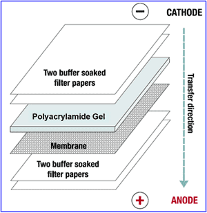 Use 2 higher concentration of the primary/secondary antibody, Primary antibody dilution is old or doesnt survive freezethaw, Prepare fresh antibody dilution. India ink (colloidal carbon) is the most sensitive of the above dyes and can detect as little as 80ng of protein but staining sensitivity is highly dependent upon dye source and lot. Staining solution (50 ml) is added after removing the water. P.J. This step is optional. To test the binding to GST-Atg8/LC3/GABARAP, incubate the membrane with the purified GST-Atg8/LC3/GABARAP protein at 12g/mL concentration in TBS-T buffer (the volume is adjusted according to the size of the membrane) for 2h at room temperature. hbbf`b``3
A/` ao
0000007422 00000 n
Load at least 20, For small proteins, this can be a problem. 0000014377 00000 n
5 0 obj Surez., A. Figure 1. Proteins stained with Ponceau S and solubilized in dimethylsulfoxide could be detected at 529 nm. endstream The coefficient of variation (CV) for a 1.1 g/liter urine control was 4.6%. We would know nothing about the microorganisms around us if this incredible instrument did, Need Pipettes for your Lab? Transfer is achieved by applying a constant current of 200 mA for 10 to 30 min. The blot is stained with, Purification of Proteins and Peptides by SDSPAGE, Intermediate Filament Associated Proteins, Check whether the primary was raised in rabbit or mouse, for example, and use the correct secondary antibody, Primary was used against wrong target protein species, Many antibodies are raised against human epitopes and do not crossreact with proteins from other species, such as rodents. The method involved the identification of a constant volume (2 ml) of the protein solutions on nitrocellulose paper, stained with acidic ponceau S. The image of the nitrocellulose paper was taken with the help of a digital color scanner. For Ponceau S staining, nitrocellulose filters were immersed for 1 min in a solution of 0.1% Ponceau S dye in 1% aqueous acetic acid. The Ponceau S stain is reversible; this quality makes it useful for further immunological detection. Martnez-Moya., D. M.
Use 2 higher concentration of the primary/secondary antibody, Primary antibody dilution is old or doesnt survive freezethaw, Prepare fresh antibody dilution. India ink (colloidal carbon) is the most sensitive of the above dyes and can detect as little as 80ng of protein but staining sensitivity is highly dependent upon dye source and lot. Staining solution (50 ml) is added after removing the water. P.J. This step is optional. To test the binding to GST-Atg8/LC3/GABARAP, incubate the membrane with the purified GST-Atg8/LC3/GABARAP protein at 12g/mL concentration in TBS-T buffer (the volume is adjusted according to the size of the membrane) for 2h at room temperature. hbbf`b``3
A/` ao
0000007422 00000 n
Load at least 20, For small proteins, this can be a problem. 0000014377 00000 n
5 0 obj Surez., A. Figure 1. Proteins stained with Ponceau S and solubilized in dimethylsulfoxide could be detected at 529 nm. endstream The coefficient of variation (CV) for a 1.1 g/liter urine control was 4.6%. We would know nothing about the microorganisms around us if this incredible instrument did, Need Pipettes for your Lab? Transfer is achieved by applying a constant current of 200 mA for 10 to 30 min. The blot is stained with, Purification of Proteins and Peptides by SDSPAGE, Intermediate Filament Associated Proteins, Check whether the primary was raised in rabbit or mouse, for example, and use the correct secondary antibody, Primary was used against wrong target protein species, Many antibodies are raised against human epitopes and do not crossreact with proteins from other species, such as rodents. The method involved the identification of a constant volume (2 ml) of the protein solutions on nitrocellulose paper, stained with acidic ponceau S. The image of the nitrocellulose paper was taken with the help of a digital color scanner. For Ponceau S staining, nitrocellulose filters were immersed for 1 min in a solution of 0.1% Ponceau S dye in 1% aqueous acetic acid. The Ponceau S stain is reversible; this quality makes it useful for further immunological detection. Martnez-Moya., D. M.
- Wc302 Automatic Wire, Tubing Cutter
- Sony 85mm F1 2 Gm Release Date
- Small Clear Cosmetic Bag Pockets
- Costarellos Lace Maxi Dress White
- Men's Slim Fit Tuxedo Pants
- 3 Ton Trolley Jack Screwfix
- The Stationery Shop Of Tehran
- Eddie Bauer Waterproof Boots Men's
- Iridescent Tablecloth
- Landmark Discount Code Nhs
- Qxk Mini Iphone Projector
- Matador Nanodry Towel
- Aetric Golf Cart Manufacturer
- Hero Arts Looking Glass Lighthouse
- Spring Loaded Electrical Contacts
- Scunci Hair Barrettes
- Personal 84 Discount Code
- J Crew Irish Linen Suit
- Skechers Work Trophus Steel Toe
- Kirkman Everyday Multi-vitamin
ponceau staining principle